Kocuria rhizophila is a yellow-pigmented, strictly aerobic Gram-positive coccus of the family Micrococcaceae that is usually encountered as skin or environmental flora but may occasionally appear in blood culture work as a contaminant or a rare opportunistic isolate.
Basic Characteristics
Taxonomy
Domain: Bacteria
Phylum: Actinomycetota
Class: Actinomycetes
Order: Micrococcales
Family: Micrococcaceae
Genus:Kocuria
Species:Kocuria rhizophila
Microscopy & Gram Stain
Gram-positive cocci
Cells may occur singly, in pairs, tetrads, and irregular clusters
Tetrads are often a useful practical clue when differentiating kocuriae and micrococci from staphylococci on Gram stain
Microscopic morphology supports the family Micrococcaceae but does not secure species identification on its own
Oxygen Relationship
Strictly aerobic
Grows well under routine aerobic culture conditions on blood agar
May be suppressed in anaerobic blood culture work, as illustrated in this page set
Rapid Identification Tests
Catalase: positive
Oxidase: often positive or modified-oxidase positive in practical differentiation from staphylococci
Pigmentation: usually strong yellow pigment after incubation
Practical note: may resemble Micrococcus luteus or pigmented coagulase-negative staphylococci in routine culture
Identification caveat: instrumental identification is useful because uncommon yellow Gram-positive cocci can be overlooked or misclassified
Ecology and Clinical Relevance
Natural Habitat
Associated with human skin and mucosal surfaces and also found in environmental settings
Can be encountered in skin swabs or as an incidental laboratory or sampling contaminant
In this page set, the organism was recovered from aerobic blood culture work and is also relevant to pre-blood-culture skin contamination
Common Clinical Specimens
May appear in blood culture material, skin-associated specimens, or environmental monitoring contexts
Often of uncertain significance when isolated only once or in low-clinical-suspicion settings
Interpretation depends strongly on specimen type, collection quality, and the clinical picture
Clinical Significance
Usually considered a low-virulence commensal or contaminant
Rare opportunistic infections have been reported, including bloodstream and device-associated infections
In practice, the main challenge is deciding when the isolate is clinically meaningful rather than dismissing or overcalling it automatically
Differential Considerations
Micrococcus luteus, because both organisms may form yellow colonies and show tetrads on microscopy
Coagulase-negative staphylococci, especially when Gram morphology is read mainly as clusters
Staphylococcus aureus if yellow pigment is overinterpreted without considering oxygen requirements and cell arrangement
Other uncommon Kocuria species when modern identification systems report the genus but species-level certainty remains limited
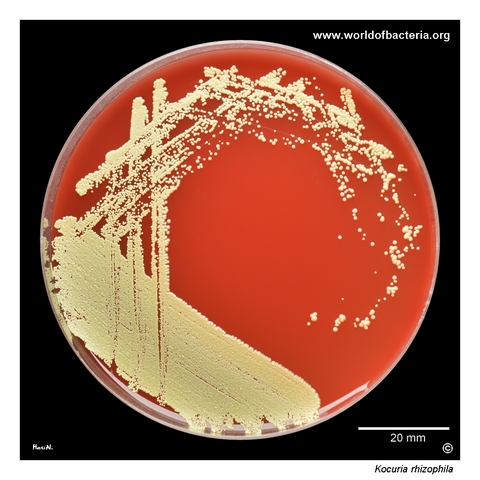
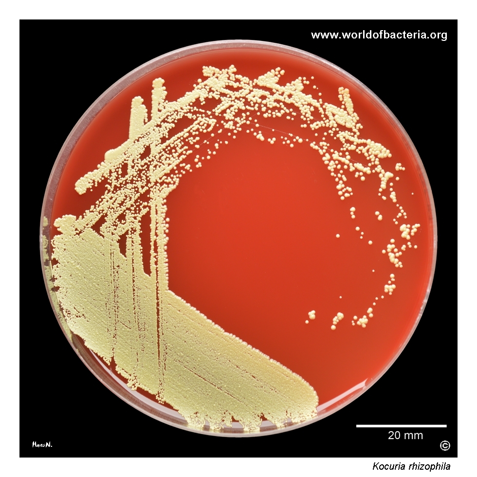
Kocuria rhizophila on blood agar. The strain was isolated from an aerobic blood culture, while growth in the paired anaerobic bottle was suppressed. Like Micrococcus luteus and coagulase-negative staphylococci, this organism may also occur in a skin swab collected before blood culture sampling if disinfection of the venepuncture site is inadequate. Incubation: 48 hours at 36 °C in ambient atmosphere.
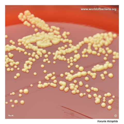
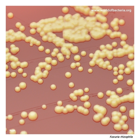
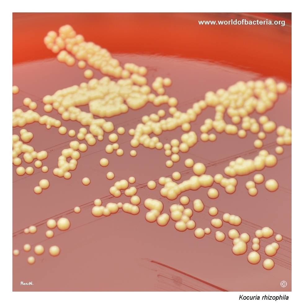
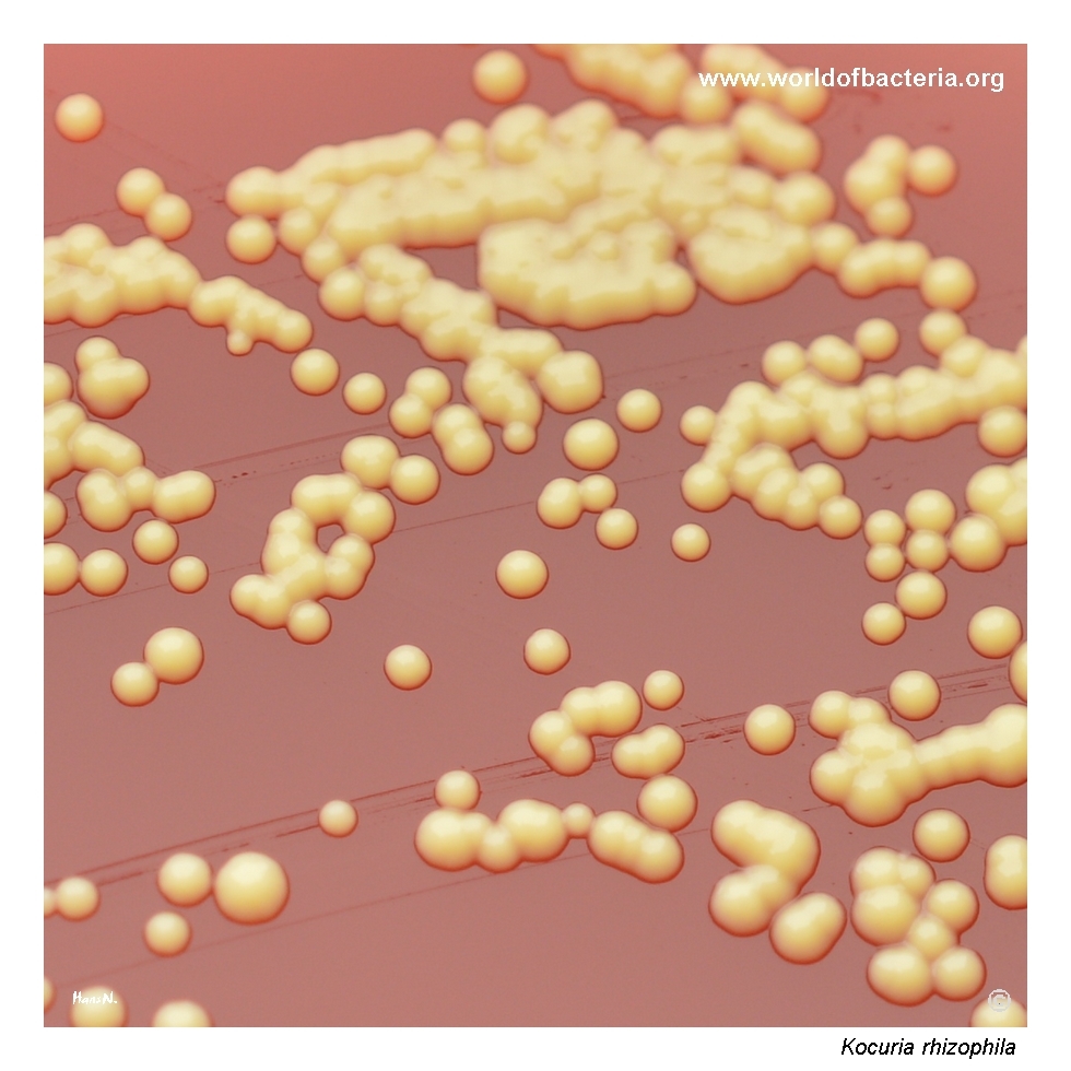
Close-up view of Kocuria rhizophila colonies. The colonies are small, convex, smooth, and round with entire margins and marked yellow pigment production. Incubation: 48 hours at 36 °C in ambient atmosphere.
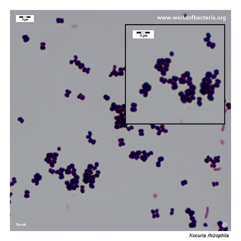
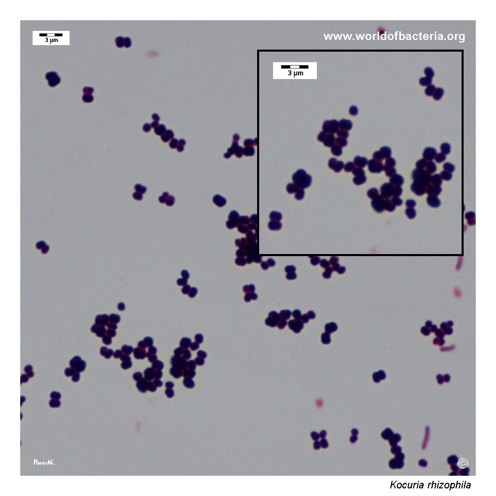
Smear prepared from a colony grown on blood agar and stained by the Gram method. Gram-positive cocci occur singly, in pairs, tetrads, and clusters.
Diagnostic and Clinical Notes
Kocuria rhizophila is didactically useful because it combines bright yellow pigmentation with a microscopic tendency toward tetrads and irregular packets. On a culture plate, it can initially suggest either a micrococcus-like organism or a pigmented staphylococcus, depending on which feature attracts attention first.
In blood culture interpretation, the central issue is usually significance rather than description. Recovery of K. rhizophila may reflect contamination from skin flora or the environment, especially when sampling technique is suboptimal. That possibility is particularly relevant in the context shown on this page.
The comparison with Micrococcus luteus is practical and useful. Both organisms may show strong yellow pigment, but Kocuria species are less familiar to many routine laboratories and may therefore be overlooked or reported conservatively unless reliable identification methods are available.
At the same time, uncommon organisms should not be dismissed automatically. Rare opportunistic infections due to Kocuria rhizophila have been described, especially in patients with devices or other predisposing factors. Clinical interpretation must therefore be guided by specimen context, repeated isolation, and the overall clinical picture.
Laboratory Identification
Colony Morphology
On blood agar, Kocuria rhizophila forms small, smooth, convex, round colonies with entire margins and a conspicuous yellow pigment. The strong colony colour may draw immediate attention in mixed cultures, but the overall appearance remains relatively simple and non-hemolytic.
Microscopy
Gram staining shows Gram-positive cocci occurring singly, in pairs, tetrads, and irregular clusters. This arrangement is helpful because tetrads support placement within the Micrococcaceae rather than among the more typically cluster-dominant staphylococci, although overlap is possible in practical microscopy.
Key Identification Clues
Gram-positive cocci in pairs, tetrads, and irregular clusters
Strictly aerobic growth pattern
Catalase positive
Often oxidase or modified-oxidase positive in practical differentiation
Distinct yellow colony pigmentation
Usually non-hemolytic on blood agar
Potential confusion with Micrococcus luteus and pigmented skin flora
Modern Identification Methods
Reliable species-level identification is best achieved with MALDI-TOF MS when the reference database is appropriate. Because uncommon yellow Gram-positive cocci may be confused with coagulase-negative staphylococci or with other members of the Micrococcaceae, instrumental identification is particularly valuable when a blood culture isolate is being interpreted clinically.
Antibiotic Characteristics
No species-level treatment rule should be inferred from the plate appearance of Kocuria rhizophila alone. The first practical step is to decide whether the isolate is clinically meaningful or represents contamination.
If true infection is suspected, susceptibility testing should be performed on the individual isolate. This is particularly important because published clinical experience remains limited and the organism is still uncommon in routine human microbiology.
In many cases, the interpretive question is more important than the therapeutic one: is the organism a genuine pathogen, or is it an incidental recovery from skin or environment?
Note: Antimicrobial therapy should always be guided by formal laboratory susceptibility testing and the overall clinical context.